Understanding the Zygomatic Bone Anatomy is essential not only for academic knowledge but also for those seeking expert care in maxillofacial surgery, trauma recovery, or cosmetic reconstruction. At Max Face Implant Center, Prof. Dr. Celal Çandırlı specializes in precision-based treatment plans that are rooted in anatomical accuracy and surgical expertise.
What is the Zygomatic Bone Anatomy?
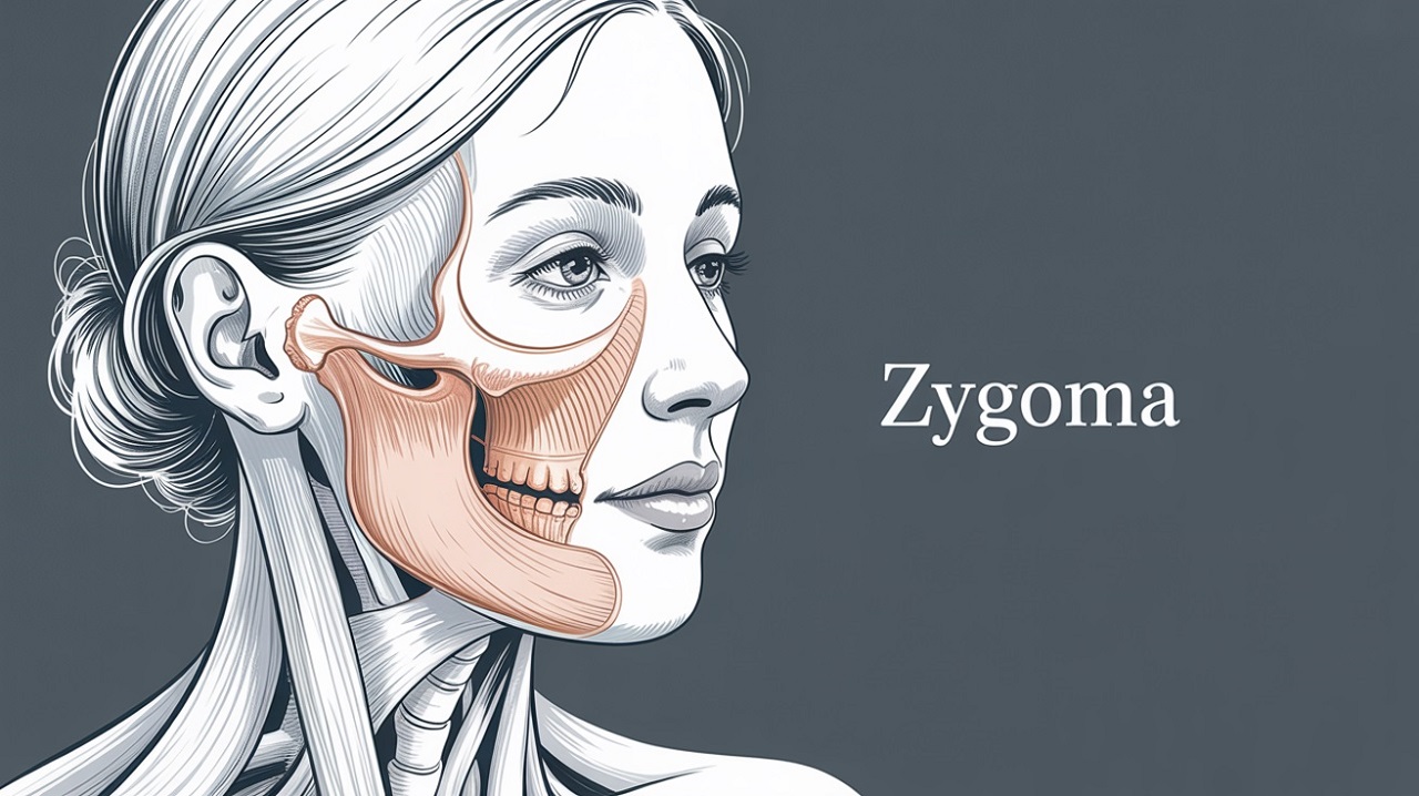
The zygomatic bone, also known as the cheekbone or malar bone, is a key structural element of the midface. Its anatomical landmarks serve as anchors for both facial aesthetics and functional architecture.
Whether you are dealing with a facial trauma, congenital defect, or seeking reconstructive surgery, understanding this anatomy is the foundation of successful treatment. That’s why patients worldwide trust Max Fax Zygoma Center for advanced diagnostics and care.
How the Zygomatic Bone Articulates with Surrounding Facial Bones?
The zygomatic bone articulates with four surrounding bones: the maxilla, temporal bone, sphenoid, and frontal bone. These articulations play a vital role in maintaining the structural integrity of the midface and serve as crucial landmarks for both anatomical understanding and surgical planning.
These bony junctions contribute significantly to the formation of the zygomatic arch and the lateral wall and floor of the orbital cavity. Together, they influence facial symmetry, support ocular structures, and enhance the overall aesthetic contour of the face.
Anatomical Landmarks of the Zygoma Processus and Borders Explained
Anatomical Landmarks of the Zygoma: Processus and Borders Explained
Each bone plays a role in nerve passage, muscle attachment, and overall structural integrity. These landmarks are critical when performing surgical reconstructions or treating zygomatic bone fractures, related with the zygomatic bone anatomy.
How is Zygomatic Process?
The zygomatic process is a bony projection that connects the zygomatic bone to neighboring bones of the skull. It is not a single structure but rather a feature found on several different bones, each contributing to the zygomatic arch and midfacial structure. Here’s a breakdown:
Types of Zygomatic Processes
Explanation
Zygomatic Process of the Maxilla
Extends laterally to articulate with the zygomatic bone, forming part of the infraorbital rim. Contributes to the anterior portion of the zygomaticomaxillary complex
Zygomatic Process of the Temporal Bone
Projects anteriorly to meet the temporal process of the zygomatic bone, forming the zygomatic arch. Important for the attachment of the masseter muscle.
Zygomatic Process of the Frontal Bone
Projects downward to articulate with the frontal process of the zygomatic bone. Forms part of the lateral orbital rim.
Temporal Process of the Zygomatic Bone
Extends posteriorly to meet the zygomatic process of the temporal bone.
The Zygomatic Arch (Structure, Function, and Clinical Significance)
The Zygomatic Arch (Structure, Function, and Clinical Significance)
The zygomatic arch forms the lateral contour of the face and provides a structural bridge between the zygoma and the temporal bone. It is vital in procedures like orthognathic surgery and midface lift interventions. Any misalignment can affect both the function and symmetry of the face.
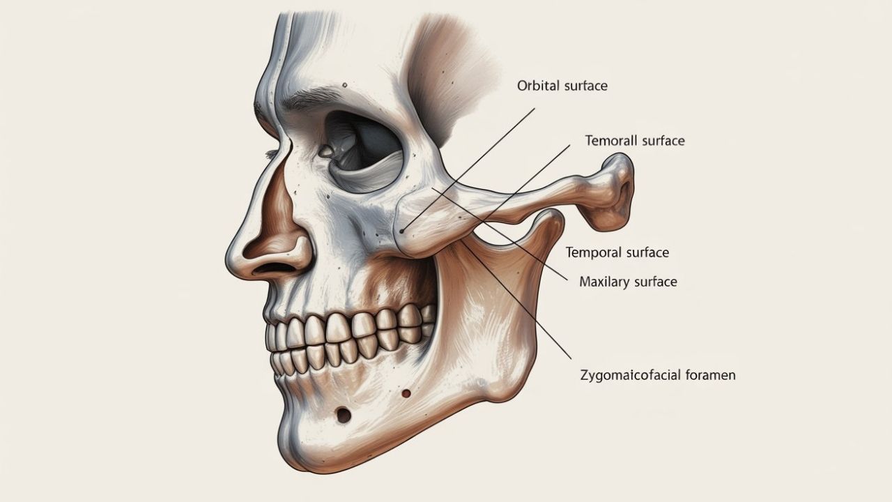
Zygomaticotemporal and Zygomaticofacial Foramina (Nerve Pathways)
Two critical foramina in the zygomatic bone anatomy—the zygomaticotemporal and zygomaticofacial foramina—allow for the transmission of sensory nerves. Proper identification of these pathways is essential during surgeries to prevent long-term sensory loss or neuropathic pain.
Zygoma’s Role in the Orbit and Eye Socket Formation
The zygomatic bone anatomy andZygoma’s Role in forming the orbital floor and lateral wall is often underestimated. Any trauma or fracture here can impact vision, eye alignment, and even neurological function. Our clinic is specialized in orbital reconstruction, ensuring both aesthetic and functional recovery.
Muscle Attachments on the Zygomatic Bone (Masseter, Zygomaticus, and More)
Several muscles insert on the zygoma, including:
Masseter (crucial for chewing)
Zygomaticus major and minor (important for facial expression)
During reconstructive or cosmetic surgeries, related with the zygomatic bone anatomy, preserving these attachments is paramount for natural movement and expression—one of the many specialties handled with expertise at Max Fax Zygoma Center.
Differences Between Left and Right Zygomatic Bones in Human Anatomy
Differences Between Left and Right Zygomatic Bones in Human Anatomy
While symmetrical by design, minor variations between the left and right zygomatic bone anatomy can exist due to trauma, genetics, or surgical history. Identifying these differences is crucial during bilateral corrective surgeries or implants.
The zygomatic bone ossifies from three centers during fetal development and continues to remodel throughout childhood. Awareness of these growth patterns aids in planning pediatric maxillofacial surgeries and anticipating post-operative changes.
Zygomatic Bone Anatomy in Humans vs. Other Mammals
Interestingly, in quadrupeds like dogs or horses, the zygomatic bone plays a different role in structural support and muscle attachment. This comparative insight enhances the understanding of biomechanics in trauma-informed surgical planning.
Clinical Importance of Zygomatic Bone Landmarks in Maxillofacial Surgery
Precise mapping of zygomatic bone landmarks is essential in various surgical procedures, including fracture fixation, facial reconstruction, orbital wall repair, and zygomatic bone reduction or augmentation. Accurate identification of these anatomical reference points ensures surgical precision, minimizes complications, and supports functional and aesthetic outcomes.
At Max Fax Zygoma Center, every surgical case is approached with advanced technology, including 3D planning, landmark-based navigation, and real-time imaging. This meticulous methodology allows our team to deliver highly personalized, safe, and effective treatments that restore both facial symmetry and confidence.Formun Altı
Why Choose Max Fax Zygoma Center?
Prof. Dr. Celal Çandırlı is internationally recognized for his expertise in Zygomatic Bone Anatomy and complex facial reconstruction. Our center provides:
Advanced imaging and modeling
Personalized treatment plans
High success rates in trauma and cosmetic cases
Book your consultation to experience evidence-based, anatomically-informed care you can trust.
From zygomatic bone fractures to advanced reconstructive surgeries, we deliver results that restore confidence and function. At Max Fax Zygoma Center, Zygomatic Bone Anatomy isn’t just a concept—it’s the cornerstone of every successful treatment.
Zygomatic Bone Anatomy – Frequently Asked Questions
What is the zygomatic bone, and why is it called the cheekbone?
The zygomatic bone (malar bone) is a key midface structure that creates the prominence of the cheeks. It also supports the lateral face contour and contributes to the orbit (eye socket), making it important for both aesthetics and function.
What bones does the zygomatic bone articulate with?
The zygomatic bone articulates with four bones: the maxilla, temporal bone, sphenoid, and frontal bone. These junctions help form the zygomatic arch and the lateral wall and floor of the orbital cavity.
What is the zygomatic arch, and what does it do?
The zygomatic arch is the bony bridge formed by the temporal process of the zygomatic bone and the zygomatic process of the temporal bone. It defines the lateral facial contour and supports chewing mechanics by providing attachment areas near the masseter region.
Which anatomical landmarks of the zygomatic bone are most important in surgery?
Key landmarks include the frontal process, temporal process, maxillary process, orbital surface, and the zygomaticofacial and zygomaticotemporal foramina. These reference points guide fracture fixation, implant placement, and orbital reconstruction while helping reduce complications.
What are the zygomaticofacial and zygomaticotemporal foramina?
These foramina are small openings in the zygomatic bone that transmit sensory nerve pathways. Careful identification during surgery helps prevent long-term numbness, altered sensation, or neuropathic pain.
How does the zygomatic bone contribute to the orbit and eye socket?
The zygomatic bone forms part of the orbital floor and the lateral orbital wall. Trauma in this region can affect eye position, alignment, and function, which is why precise orbital assessment and reconstruction planning are essential.
What is the “zygomatic process,” and is it the same on every bone?
The zygomatic process refers to a bony projection present on multiple bones, not a single identical structure. For example, the temporal bone has a zygomatic process that helps form the arch, while the maxilla and frontal bone also have zygomatic processes that connect to the zygoma.
Which muscles attach to the zygomatic bone?
Important attachments relate to facial expression and chewing, including the zygomaticus major/minor (smile and expression dynamics) and the masseter region functionally associated with the zygomatic arch. Preserving anatomical relationships supports natural movement and balanced facial outcomes.
Can differences between the left and right zygomatic bones be normal?
Yes, mild asymmetry is common due to genetics, development, or subtle lifestyle and muscular factors. However, noticeable asymmetry can also follow trauma or prior surgery and may be evaluated in planning implants or bilateral corrections.
How does the zygomatic bone develop and change over time?
The zygomatic bone ossifies from multiple centers during fetal development and continues remodeling throughout childhood. Understanding growth patterns is especially important in pediatric planning and in anticipating long-term structural changes.
Why is zygomatic bone anatomy important in maxillofacial trauma care?
Zygomatic fractures can impact facial symmetry, chewing function, and orbital integrity. Anatomical accuracy helps surgeons restore the zygomaticomaxillary complex and protect nerve pathways, improving both functional recovery and facial contour.
How is zygomatic anatomy used for implant or reconstruction planning?
Landmark-based evaluation supports precise implant positioning and balanced projection in cosmetic or reconstructive cases. Advanced planning methods (such as 3D modeling) help align the midface, support the orbit, and optimize predictable outcomes.
What symptoms can occur after a zygomatic fracture involving the orbit?
Patients may experience swelling, facial flattening, pain with chewing, numbness, or changes around the eye such as misalignment or visual disturbance. A specialized evaluation helps determine whether orbital floor or lateral wall reconstruction is necessary.
Is the zygomatic bone anatomy the same in humans and other mammals?
The general concept of a cheekbone region exists across many mammals, but its shape and biomechanical role can differ significantly. In quadrupeds, the zygomatic region often reflects species-specific chewing demands and skull mechanics.
When should someone seek specialist evaluation for zygomatic bone issues?
If there is facial trauma, suspected fracture, persistent numbness, asymmetry, or functional issues involving chewing or the eye area, a specialist assessment is recommended. A maxillofacial surgeon can evaluate bone alignment, orbital involvement, and nerve pathways using advanced imaging.


 Why Choose Max Fax Zygoma Center?