Benefits of 3D Imaging for Zygomatic Implant Surgery Planning
3D imaging plays an important role in zygomatic implant surgery planning. It allows clinicians to examine the upper jaw and cheekbone structures in detail before any procedure begins.Traditional two-dimensional radiographs often leave gaps in understanding bone volume and nerve pathways. Three-dimensional scans reduce these limitations and support more accurate decisions during digital planning.This article explains how 3D imaging zygomatic implants benefit from improved visualization, how surgery planning accuracy increases, and why ct scan benefits matter for precise implant placement.
What 3D Imaging Shows Before Surgery
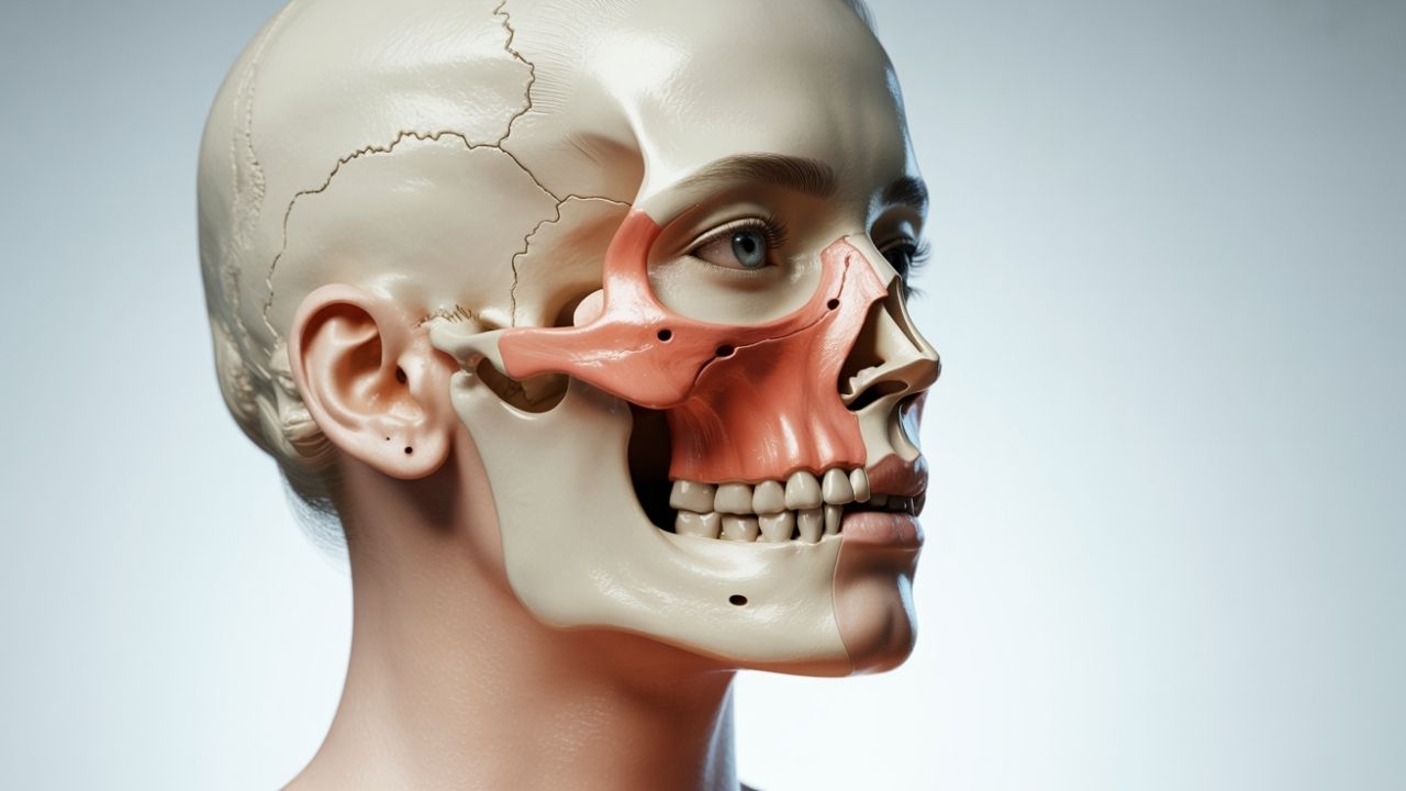
Three-dimensional scans display bone height, width, and density in multiple planes. They also reveal the relationship between the maxillary sinus and the zygomatic bone.Clinicians can rotate and section the images to study anatomical variations that may affect implant angulation. This level of detail supports safer planning for each individual case.
Comparison of Planning Methods
| Method | Key Features | Limitations |
|---|
| Traditional 2D X-ray | Quick and widely available | Limited depth perception and bone detail |
| CT or CBCT Scan | Full 3D view of bone and sinuses | Higher radiation than basic radiographs |
| Digital Planning Software | Virtual implant placement and measurements | Requires compatible scan data |
How Accuracy Improves with Digital Tools
Digital planning software lets the team simulate different implant positions on the screen. Measurements of bone thickness and implant length can be taken directly from the images.Precise implant placement becomes more predictable when the surgeon can visualize the final position before entering the operating room. This reduces the chance of unexpected findings during surgery.
Points Patients Should Discuss
- Which imaging method will be used and why
- How radiation exposure is kept as low as possible
- What information the scan provides about individual anatomy
- Whether additional scans will be needed later
Sık Sorulan Sorular
Does 3D imaging replace the need for clinical examination?
No. 3D imaging supports planning but cannot replace a thorough in-person evaluation by a qualified professional.
How does ct scan benefit zygomatic implant cases?
CT scans provide detailed views of bone quality and sinus anatomy that help determine whether zygomatic implants are suitable for a specific patient.
Is radiation exposure a concern with 3D scans?
Modern cone-beam systems use lower doses than traditional medical CT, yet the decision to use any scan remains individual and should be discussed with the clinician.
Can digital planning guarantee exact implant position?
Digital planning improves surgery planning accuracy but cannot guarantee outcomes. Actual placement still depends on surgical conditions and healing responses.
Who decides if 3D imaging is necessary?
The treating specialist evaluates each case based on medical history, clinical findings, and available imaging options.
Are there alternatives to 3D imaging for planning?
Some simpler cases may rely on panoramic radiographs, but complex zygomatic implant situations often benefit from three-dimensional data for better visualization.
Conclusion
3D imaging supports more informed zygomatic implant surgery planning by showing bone structures and anatomical relationships clearly. Every patient’s situation differs, so suitability for any imaging or surgical approach requires personal assessment by a qualified health professional.
Expert Information
This content has been prepared for general informational purposes in the field of oral, dental and maxillofacial surgery. Prof. Dr. Celal Çandırlı’s more than 20 years of professional experience and training at England NHS Poole Hospital and Newcastle Freeman Hospital contribute to the scientific and clinical presentation of the topic.
This content is for general information purposes only and does not replace diagnosis or treatment. For complaints related to oral, dental and jaw health, consult your dentist or relevant specialist healthcare professional.